Design.

- To create a 3D printed liver, Polycaprolactone (PCL) coated with liver extracellular matrix (ECM) will be used as the “ink” in the 3D printer. PCL was chosen due to its biocompatibility and biodegradability. PCL is also widely available and cost effective. Its mechanical strength and porosity can be adjusted [14].
- The scaffold will be designed on a software that can be sent to a 3D printer. It will feature 8 different pieces that mirror the Couinaid segments to help with vascularization, as each Couniaid segment in the liver can function independently and has their own vascular inflow and outflow [17].
- To vascularize the liver, the 3D printing technique that we will be using is called extrusion. This technique will precisely position cells in layers with x, y, and z coordinates. The resulting scaffold will have vascular networks with perusable channels [18].
- Lastly, hepatoblasts (which make up the majority of the liver) will be inserted into the scaffold with a hydrogel mixture of gelatin, fibrinogen, hyaluronic acid, and glycerol mixed into DMEM [19].
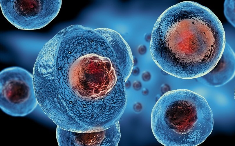
Stem Cells
While we had contemplated using donated cells from the ACLF patient to use in the liver for a better organ match, using stem cells would allow us to create livers ahead of time. Because ACLF progresses so fast, time is not on our side. With this way, once patients are diagnosed with ACLF, this 3D printed liver can already an option for them.
Embryonic stem cells will be soaked in ECELS Media until hepatoblast phenotypes are displayed. ECELS media helps prolong the display of hepatoblast phenotypes and stabilizes this process when compared to a previous study done under the same conditions. Once hepatoblast phenotypes are displayed, they can be exposed to hepatocyte developmental cues which help the hepatoblasts differentiate into mature hepatocytes [11].
Contents of ECELS Media: basal media supplemented with insulin, transferrin, sodium selenite, epidermal growth factor, glycogen synthase kinase 3 inhibitor, transforming growth factor β receptor inhibitor, lysophosphatidic acid, and sphingosine 1-phosphate [11].
Hepatocyte Developmental Cues: For 2 weeks, hepatocytes will sit in HOD media (hepatocyte Growth Factor, dexamethasone, and Oncostatin M) and is treated with 2 μM TGF-β receptor inhibitor (SB431542) and 1 μM γ-secretase inhibitor (RO4929097) before they become hepatoblasts [11].
Preservation
For most organ transplantations, organs are preserved at temperatures above-zero by using a process known as cryogenic cooling. In this process, tissues or cells are “frozen” in temperatures above-zero. The sample, however, is only preserved for around four to twelve hours. [20] Vitrification is another method of preserving tissues and cells without ice, but can only be kept for around the same time. [21] An ex vivo machine perfusion system, shown in the image below, can be used to preserve tissues and organs at -6 degrees Celsius for up to 96 hours without ever freezing the organ tissue itself. [22] In our design, we will follow a similar method.

Alternative Methods
There are two alternative methods that we can use in our design. The first method is using Carbon Nanotubes, a type of scaffold that works well with stem cells, but has not been used with 3D printing before. [12]
The second method is by using Alginate. Alginate is a type of 3D printed scaffold with high biocompatibility and biodegradability. However, alginate is biologically inert and does not work well with stem cell differentiation. [23]
Testing
To test the scaffolds in our design, we will use scaffolds implanted in rats in order to see how the engineered tissue and the scaffold hold up when implanted in an organism. [24] In order to test the engineered liver’s function, itself, we can use nuclear medicine. Small amounts of a radioactive substance can be injected into a patient’s bloodstream. Devices could then detect the radiation in the blood as it flows throughout the liver, testing its functionality. [25]
![]()
References:
11. Zhang, M., Sun, P., Wang, Y., Chen, J., Lv, L., Wei, W., . . . Li, W. (2015). Generation of Self-Renewing Hepatoblasts From Human Embryonic Stem Cells by Chemical Approaches. Stem Cells Transl Med, 4(11), 1275-1282. doi:10.5966/sctm.2015-0051
12.Brunner, E.W., et al., Growth and proliferation of human embryonic stem cells on fully synthetic scaffolds based on carbon nanotubes. ACS Appl Mater Interfaces, 2014. 6(4): p.2598-603.
14. Mirdamadi, E. S., Kalhori, D., Zakeri, N., Azarpira, N., & Solati-Hashjin, M. (2020). Liver Tissue Engineering as an Emerging Alternative for Liver Disease Treatment. Tissue Eng Part B Rev, 26(2), 145-163. doi:10.1089/ten.TEB.2019.0233
17. Sibulesky, L. (2013). Normal liver anatomy. Clin Liver Dis (Hoboken), 2(Suppl 1), S1-S3. doi:10.1002/cld.124
18. Jafarkhani, M., et al., Bioprinting in Vascularization Strategies. Iran Biomed J, 2019. 23(1): p. 9-20.
19. Kang, H. W., Lee, S. J., Ko, I. K., Kengla, C., Yoo, J. J., & Atala, A. (2016). A 3D bioprinting system to produce human-scale tissue constructs with structural integrity. Nat Biotechnol, 34(3), 312-319. doi:10.1038/nbt.3413
20. Liu, D. and F. Pan, Advances in cryopreservation of organs. J Huazhong Univ Sci Technolog Med Sci, 2016. 36(2): p. 153-161.
21. Fahy, G.M. and B. Wowk, Principles of cryopreservation by vitrification. Methods Mol Biol, 2015. 1257: p. 21-82.
22.Bruinsma, B.G., et al., Supercooling preservation and transplantation of the rat liver. Nat Protoc, 2015. 10(3): p. 484-94.
23.Mazza, G., et al., Liver tissue engineering: From implantable tissue to whole organ engineering. Hepatol Commun, 2018. 2(2): p. 131-141.
24. Khorramirouz, R., et al., A novel surgical technique for a rat subcutaneous implantation of a tissue engineered scaffold. Acta Histochem, 2018. 120(3): p. 282-291.
25. Bennink, R.J., et al., Liver function testing with nuclear medicine techniques is coming of age. Semin Nucl Med, 2012. 42(2): p. 124-37.
Figure 12: https://www.spandidos-publications.com/10.3892/etm.2016.3206
Figure 13: https://3dprint.com/7729/3d-print-organs-vascular/
Figure 14: https://pubs.rsc.org/en/content/articlelanding/2019/tb/c9tb00485h#!divAbstract
Figure 15: https://biologydictionary.net/wp-content/uploads/2018/03/Embryonic-Stem-Cell.jpg
Figure 16: https://www.cbc.ca/news/canada/british-columbia/new-transplant-tech-extends-organ-life-outside-body-1.5248255
Figure 17: https://neurophilosophy.wordpress.com/2006/03/17/123/
Figure 18: https://www.researchgate.net/figure/Three-dimensional-printed-alginate-scaffold-of-14-layers-224-mm-in-height-A-Top_fig2_320793216
Figure 19: https://chernobylguide.com/nuclear_medicine/