Research overview
Our laboratory is interested in the basic molecular mechanisms of how the vertebrate body plan is established during embryogenesis, as well as broad questions regarding the genetic and cellular properties of stem cells and cancer metastasis. We use zebrafish embryos as our model system, which are ideally suited for studying in vivo stem cell and cancer biology. Zebrafish embryos develop outside the mother and are transparent, allowing for the direct visualization of individual stem cells and differentiated tissues. Zebrafish are amenable to genetic manipulations including targeted loss of function mutations and stable transgenesis. Additionally, human cancer cells or cells from early zebrafish embryos can be transplanted into host embryos for mosaic analysis. These traits allow us to manipulate genes and signaling pathways in a temporal manner at the single cell level, and then directly visualize cell behavior and differentiation in an intact living embryo. This is not possible in any other vertebrate model system.
Using neuromesodermal progenitors as a simple in vivo model of stem cell biology
All vertebrate embryos develop in an anterior to posterior progression, with the head forming first and the rest of the body extending away from the head. A group of cells in an embryonic structure called the tailbud, fuel this process of posterior growth by contributing new cells to the extending body axis. These cells, now called neuromesodermal progenitors (NMPs), exhibit extreme plasticity, and their identification has caused a paradigm shift in how we think about germ layer formation and the basic principles of vertebrate body formation. NMPs make a germ layer decision when they give rise to either neural ectoderm (spinal cord) or mesoderm (muscle, blood vessels, and other tissues). This property o f NMPs indicates that germ layer formation continues after gastrulation in the tailbud, and that spinal cord and certain mesodermal cells are more closely related to each other than to other cells within the same germ layer. The imaging and experimental accessibility of the zebrafish tailbud, combined with the stem cell-like properties of the NMPs, provide unparalleled opportunities to study stem/progenitor cell questions in an in vivo context. We currently have several major projects utilizing NMPs as in vivo experimental model.
Cell cycle control of invasive behavior during development and cancer metastasis
This project is a collaboration with the laboratory of David Matus, and is founded upon an experimental observation made in the model roundworm, C. elegans, that cell invasion and cell division are mutually exclusive behaviors. In other words, a cell cannot simultaneously invade and divide. This functional link between cell cycle arrest and invasive behavior has not been directly made before, although in a variety of cancers there exists correlative data suggesting that tumor cells become less proliferative during invasion. Our lab is using high-resolution imaging at single-cell and subcellular resol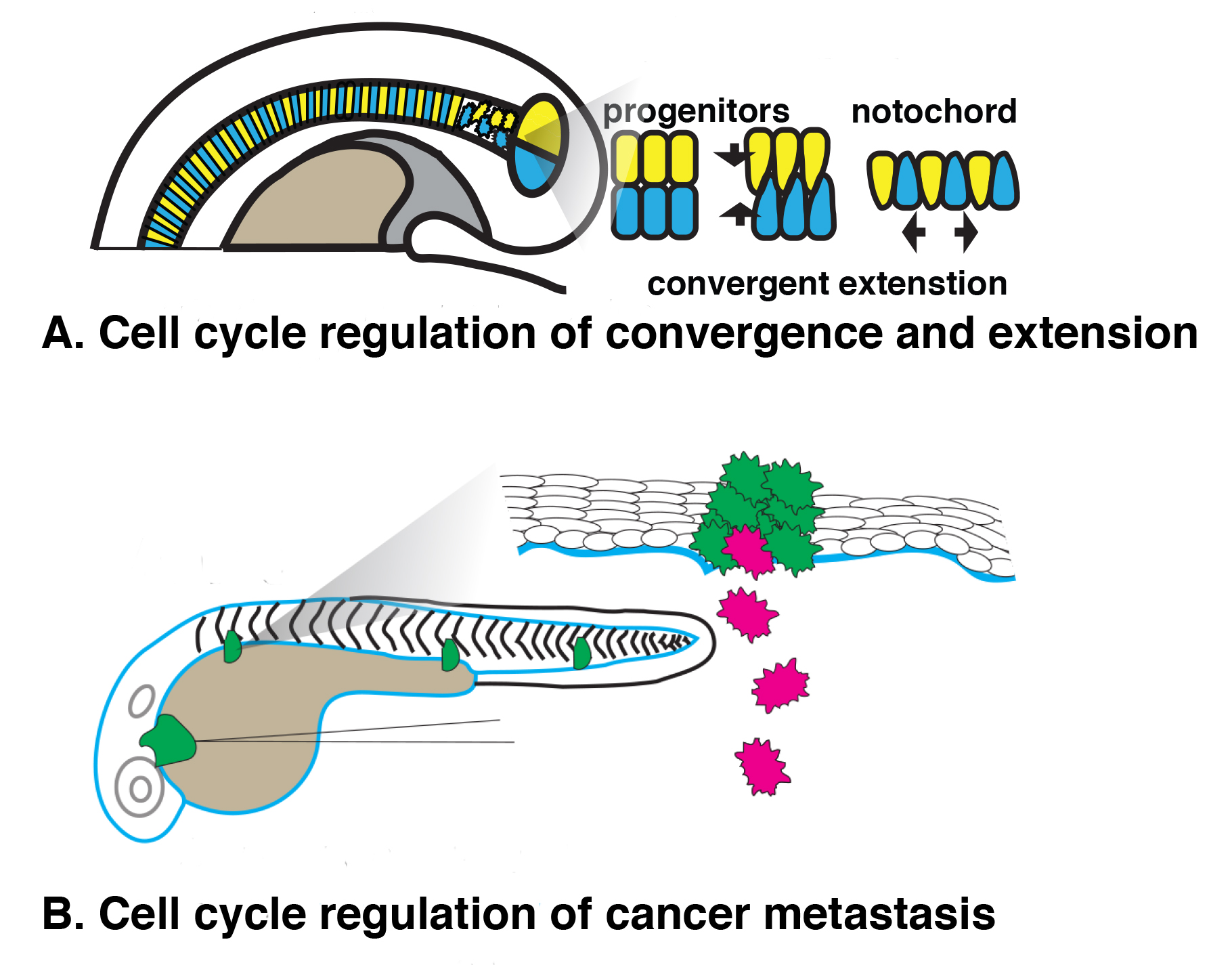 ution paired with powerful genetic and functional genomic approaches to explore the relationship between cell cycle arrest and invasive behavior during development and cancer progression. In one model, we are examining notochord differentiation during development, where a specialized set of progenitor cells undergo an invasive event called convergent extension, a cellular behavior that is also used by cancer cells during metastasis. In a direct examination of cancer cell invasion, we are using a zebrafish xenograft system that allows us to observe human cancer cell behavior in an in vivo environment.