Stony Brook University
Stony Brook, NY
Medical Physics | 2019 – Current
Optimization of Digital Breast Tomography Methods
My current graduate research under Dr. Wei Zhao focuses on the improvement of breast imaging techniques. Mammography trials are simulated through In Silico imaging, which allows us to digitally simulate a full-field mammography exam on a digital breast phantom without unnecessary radiation exposure. In silico techniques allow a greater control over the trial dosage, angular spread, breast density, micro calcification size, and other important factors. Four-alternative forced choice (4AFC) tests are used to determine the micro calcification visibility of our collected data. Our end goal is to produce nearly noise-less images that can clearly show micro-calcifications with the technology we presently hold, therefore resulting in accurate and early breast cancer detection.

Biomedical Engineering | Summer 2018
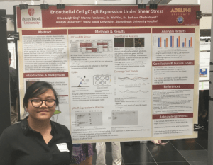
Endothelial Cell qC1qR Expression Under Shear Stress
My NSF funded REU was through the Center of Inclusive Education (CIE). Under Dr. Wei Yin, I was a part of a pathology research that focused on protein sheering and the effects inflammation over time. While this research was unrelated to my general interests, I still learned a great deal about wet-lab procedure, lab regulations, and other processes that would be atypical in physics and statistical research. This research opportunity also led me to apply and attend Stony Brook University. My abstract is re-quoted below:
Angioedema is an inflammatory disorder characterized by recurrent severe swelling in the limbs, face, intestinal tract, and airways. Mutations in the SERPING1 gene decrease production of C1 inhibitor–a protein that normally inhibits inflammation. Without C1-INH, an abundance of bradykinin and gC1q-Receptors cause fluid leaking from endothelial cells into tissue causing swelling. The effects of shear stress on gC1qR expression are unknown in angioedema, but we believe that shear stress will increase gC1qR expression. We applied physiological and pathological shear stress that would be found in microvasculature to endothelial cells through an in vitro cone and plate shearing device. An ELISA was conducted after shearing to measure gC1qR expression. Cells were stained with fluorescent tags to visualize morphology. Results show that gC1qR expression is increased under physiological stress, but is unchanged under pathological stress. With this evidence, we hope to quantify gap junction size and test peptides that block interactions with gC1qR.
Adelphi University | NYU Winthrop Hospital
Garden City, NY | Mineola, NY
Medical Physics | 2017 – 2019
Landauer Microstar II in Diagnostic Dose Optimization
My undergraduate research was under NYU Winthrop Hospital’s Medical Physics team. I was under the guidance of Susanna Reis, M.S. and mentored by Prof. Matthew Wright. This was my first research project and greatly influenced my current graduate studies. My abstract is re-quoted below:
The purpose of this paper is to explore the usefulness of the Landauer Microstar II nanoDot dosimeter. When advertised, it was meant to be a utilitarian device with an extensive energy range that would be usable for both imaging physics and radiation therapy. Despite its ambitious description, nanoDots are virtually unused throughout all forms of clinical physics. By the end of my study, I concluded that nanoDots are completely unusable in health care procedures because of their inaccessibility, tedious nature, and non-ergonomic design. Despite its shortcomings, nanoDots still find extensive use in radiation research.